post

When Cells Rebel: the dark side of evolution

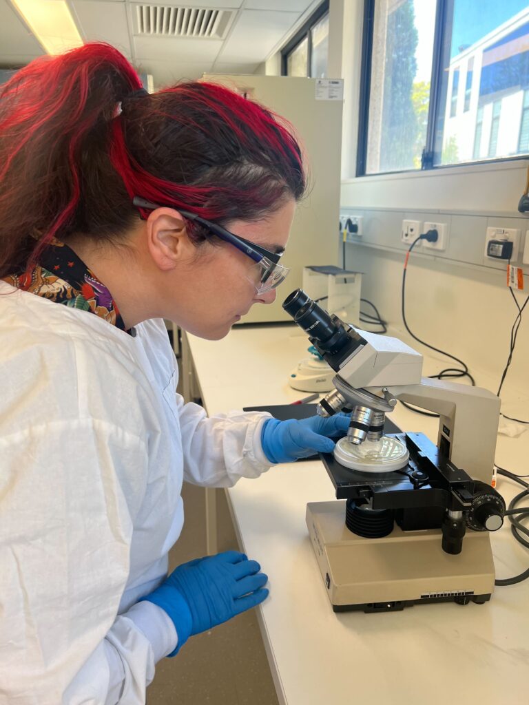
by Patra Petrohilos (PhD Student)

I love dystopian horror. I love to relish in the thrill of disgust from the comfort of safety – a comfort bolstered by the knowledge that such grotesquerie could never actually happen in real life. Zombies don’t exist. Monsters aren’t trying to escape from the underworld. Cancer isn’t contagious. Actually, maybe scratch that last one. . .

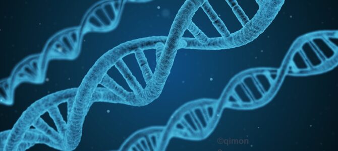
You see, nature may not have the imagination of Stephen King, but it does have something even more powerful in its arsenal: mutations. Mutations are to evolution what creativity is to horror writers – the raw material that allows them to conjure up new and wondrous forms. From the most beautiful (buttercups, butterflies, butter yellow bumblebees) to the most horrific (flesh eating bacteria, pandemic inducing viruses, cancer cells).

Evolution favours the fittest individuals, be they butterflies or bacteria. In this case, “the fittest” just means the ones that are most successful at reproducing. If we are talking about koalas, reproduction means making cute little baby koalas. Everyone likes those. But when we’re talking about cancer cells, reproduction means growing and spreading and killing one’s host. Nobody likes that. Even the cancer cells probably wouldn’t like it – because killing their host also means killing themselves in the process. Kind of like a suicide bomber without the political motivation. But evolution is blind to morality and selects for the cute little baby koalas and murderous cancer cells equally – whatever is most efficient at making more copies of itself. Survival of the fittest.

Mutations are constantly arising in nature. Sometimes these make more successful versions of things, sometimes less successful. It’s a bit of a trial-and-error process. And somewhere in that trial-and-error process, a handful of cells have stumbled across the secret to become the most successful cancer cells ever. Super-cancers! How? By finding a sneaky way around that whole unfortunate dying-when-your-host-dies bit.

They do this by taking a leaf out of the life history book of parasites. Like cancer cells, many parasites are reliant on a host to survive. But unlike cancer cells, many parasites have the power to survive the death of their host by simply finding a new host – a power that evolution has also bestowed upon these super-cancers.

Yes, nature has managed to take one of the most awful diseases known to humanity and done perhaps the only thing that could make it worse. It has made it contagious.

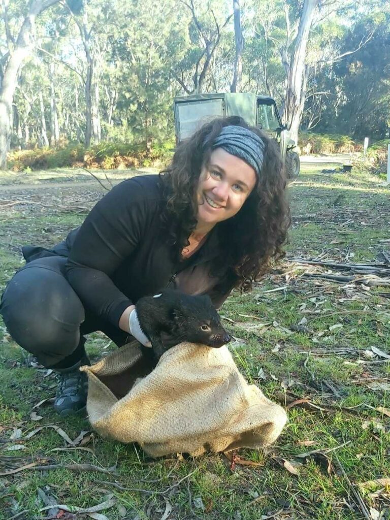
Thankfully, such contagious super-cancers are mercifully rare and none of them affect humans (yet). But the rest of the animal kingdom has not fared quite so well. Leukaemia cells drift through the sea like hidden assassins, spreading from one unsuspecting clam to the next. Dogs can get mushroom shaped tumours on their penises from sex with a poorly chosen partner. And one of our most iconic Australian animals, the Tasmanian devil, is at risk of extinction from not only one but two contagious cancers (creatively named Devil Facial Tumour Disease 1 and Devil Facial Tumour Disease 2). Sometimes lightning really does strike twice.

The good news is, this is where we come in. By researching Devil Facial Tumour Disease – one of the most uniquely horrifying and bizarre diseases to ever arise – we aim to understand how it works, how it spreads, how it evolves and, hopefully one day, how we can stop it.

Follow me for more fun and uplifting facts about the animal world!

Author:

Patra Petrohilos

Patra Petrohilos (PhD Student) is researching the evolution of devil facial tumour disease (DFTD). By investigating anticancer properties of naturally occurring peptides, she is aiming to identify novel agents with therapeutic potential against DFTD. Patra Petrohilos is a PhD student with the Australian Research Council Centre of Excellence for Innovations in Peptide and Protein Science (CIPPS). Follow their exciting research at https://cipps.org.au.

Remnant kenngoor (Phascogale calura) retain genetic connectivity and genetic diversity in a highly fragmented landscape

Type: Journal Article

Reference: de Visser, R. S., Hall, M., Ottewell, K., Pierson, J. C., Sanders, A., Friend, J. A., Berry, L., Hogg, C. & Catullo, R. A. (2024). Remnant kenngoor (Phascogale calura) retain genetic connectivity and genetic diversity in a highly fragmented landscape. Conservation Genetics, 1-15. https://doi.org/10.1007/s10592-024-01603-z

Abstract

Kenngoor (Phascogale calura) persist in < 1% of their original distribution, occupying highly fragmented remnant habitat in south-west Western Australia, with very little known of the genetic diversity of the remaining wild populations. Recently, the species has been translocated to managed reserves to improve its conservation. Understanding genetic structure and patterns of genetic diversity is crucial to inform conservation translocations for species recovery. This study aims to (1) assess genetic structure and genetic diversity across remaining wild locations, (2) assess long-term genetic outcomes of a mixed-source wild-to-wild translocation, and (3) estimate global effective population size. We genotyped 209 samples from 13 locations of fragmented remnant habitat using reduced representation sequencing. An isolation by distance model best explained genetic structure across the survey areas, with evidence of fine scale divergence of two northern locations. Allelic richness and autosomal heterozygosity measures indicated that diversity is spread uniformly across locations, and no locations showed signs of inbreeding or strong genetic drift. The mixed-source translocation has retained the diversity of the wider species ten years post-translocation. Overall, our results suggest that connectivity between survey areas has largely been maintained and that no location has substantially lower genetic diversity, despite the highly fragmented nature of remnant kenngoor habitat. Future translocations should aim to represent a mixture of genetically divergent locations to maintain the diversity present at the species level. Ongoing conservation management will be required to ensure the long-term viability of the species in this fragmented landscape.

Dating in the Dark: Elevated Substitution Rates in Cave Cockroaches (Blattodea: Nocticolidae) Have Negative Impacts on Molecular Date Estimates

Type: Journal article

Reference: Toby G L Kovacs, James Walker, Simon Hellemans, Thomas Bourguignon, Nikolai J Tatarnic, Jane M McRae, Simon Y W Ho, Nathan Lo, Dating in the Dark: Elevated Substitution Rates in Cave Cockroaches (Blattodea: Nocticolidae) Have Negative Impacts on Molecular Date Estimates, Systematic Biology, Volume 73, Issue 3, May 2024, Pages 532–545, https://doi.org/10.1093/sysbio/syae002

Abstract

Rates of nucleotide substitution vary substantially across the Tree of Life, with potentially confounding effects on phylogenetic and evolutionary analyses. A large acceleration in mitochondrial substitution rate occurs in the cockroach family Nocticolidae, which predominantly inhabit subterranean environments. To evaluate the impacts of this among-lineage rate heterogeneity on estimates of phylogenetic relationships and evolutionary timescales, we analyzed nuclear ultraconserved elements (UCEs) and mitochondrial genomes from nocticolids and other cockroaches. Substitution rates were substantially elevated in nocticolid lineages compared with other cockroaches, especially in mitochondrial protein-coding genes. This disparity in evolutionary rates is likely to have led to different evolutionary relationships being supported by phylogenetic analyses of mitochondrial genomes and UCE loci. Furthermore, Bayesian dating analyses using relaxed-clock models inferred much deeper divergence times compared with a flexible local clock. Our phylogenetic analysis of UCEs, which is the first genome-scale study to include all 13 major cockroach families, unites Corydiidae and Nocticolidae and places Anaplectidae as the sister lineage to the rest of Blattoidea. We uncover an extraordinary level of genetic divergence in Nocticolidae, including two highly distinct clades that separated ~115 million years ago despite both containing representatives of the genus Nocticola. The results of our study highlight the potential impacts of high among-lineage rate variation on estimates of phylogenetic relationships and evolutionary timescales.

A chromosome-level genome assembly for the dugong

Type: Journal Article

Reference: Dorothy Nevé Baker, Linelle Abueg, Merly Escalona, Katherine A Farquharson, Janet M Lanyon, Diana Le Duc, Torsten Schöneberg, Dominic Absolon, Ying Sims, Olivier Fedrigo, Erich D Jarvis, Katherine Belov, Carolyn J Hogg, Beth Shapiro, A chromosome-level genome assembly for the dugong (Dugong dugon), Journal of Heredity, Volume 115, Issue 2, March 2024, Pages 212–220, https://doi.org/10.1093/jhered/esae003

Abstract

The dugong (Dugong dugon) is a marine mammal widely distributed throughout the Indo-Pacific and the Red Sea, with a Vulnerable conservation status, and little is known about many of the more peripheral populations, some of which are thought to be close to extinction. We present a de novo high-quality genome assembly for the dugong from an individual belonging to the well-monitored Moreton Bay population in Queensland, Australia. Our assembly uses long-read PacBio HiFi sequencing and Omni-C data following the Vertebrate Genome Project pipeline to reach chromosome-level contiguity (24 chromosome-level scaffolds; 3.16 Gbp) and high completeness (97.9% complete BUSCOs). We observed relatively high genome-wide heterozygosity, which likely reflects historical population abundance before the last interglacial period, approximately 125,000 yr ago. Demographic inference suggests that dugong populations began declining as sea levels fell after the last interglacial period, likely a result of population fragmentation and habitat loss due to the exposure of seagrass meadows. We find no evidence for ongoing recent inbreeding in this individual. However, runs of homozygosity indicate some past inbreeding. Our draft genome assembly will enable range-wide assessments of genetic diversity and adaptation, facilitate effective management of dugong populations, and allow comparative genomics analyses including with other sirenians, the oldest marine mammal lineage.

Translating genomic advances into biodiversity conservation

Type: Journal Article

Reference: Hogg, C.J. Translating genomic advances into biodiversity conservation. Nat Rev Genet (2023). https://doi.org/10.1038/s41576-023-00671-0

Abstract

A key action of the new Global Biodiversity Framework is the maintenance of genetic diversity in all species to safeguard their adaptive potential. To achieve this goal, a translational mindset, which aims to convert results of basic research into direct practical benefits, needs to be applied to biodiversity conservation. Despite much discussion on the value of genomics to conservation, a disconnect between those generating genomic resources and those applying it to biodiversity management remains. As global efforts to generate reference genomes for non-model species increase, investment into practical biodiversity applications is critically important. Applications such as understanding population and multispecies diversity and longitudinal monitoring need support alongside education for policymakers on integrating the data into evidence-based decisions. Without such investment, the opportunity to revolutionize global biodiversity conservation using genomics will not be fully realized.

Sydney Science in Instagram: Meet PhD Student Holly Nelson

Meet PhD student Holly Nelson. Her research with the USYD Australian Wildlife Genomics group and NSW Department of Planning and Environment focuses on using genomic data to help provide tools for the management of threatened species, especially the critically endangered Bellinger River snapping turtle.

Watch the full video here: https://www.instagram.com/sydney_science/reel/Cw1FG6-hhD8/

post

How to catch a Tasmanian devil

by Dr Elspeth McLennan (Post-doc)

Tasmanian devils are nocturnal. We set traps during the day and overnight curious devils will come to investigate. The traps we use are made of strong PVC plastic fashioned into a cylinder with a spring trap door (see cover picture). The traps are baited with a devil’s favourite treat, a piece of fresh lamb or wallaby. The meat is tied onto the end of a string, fed through the trap, and tied to a pin which holds the door open.

Tasmanian devil inside trap

When a devil comes investigating the smell of the meat, they walk to the end of the trap and take the bait. When the meat is pulled and eaten, the pin holding the door open is pulled free and the door swings shut. A second pin slides forward as the door closes and locks it. Devils spend the night in a cosy enclosed space with a full belly. The field biologists begin checking the traps as soon as the sun is up. As its daylight, we often find devils snoozing in their traps.

To perform a health check on the devil, we place a hessian sack over the opening of the trap, gently tilt the trap and the devil slides forward into the sack. The sack is used to keep the devil’s eyes covered to keep them calm making them easier to handle while we check them over. We take their weight, check their body condition, look for wounds and record pouch young in females. For populations suffering from devil facial tumour disease (DFTD), the disease status of each animal is also recorded. Once the devil has been processed, they are released. On a single trapping trip, we will often see the same devils a few times. The free food and somewhere to crash is clearly a good draw.

Author

Dr Elspeth McLennan

Dr Elspeth McLennan (Post-doc) is working the on the Koala Genome Survey, investigating both neutral and functional diversity across the koala’s range to better understand the impacts of a changing climate. Elspeth has expertise in conservation genetics and using translocation and assisted colonisations as a conservation management tool.

DNA methylation networks underlying mammalian traits

Type: Journal article

Reference: Haghani, A., et al. (2023). DNA methylation networks underlying mammalian traits. Science, 381(6658), eabq5693. https://doi.org/doi:10.1126/science.abq5693

Abstract

Using DNA methylation profiles (n = 15,456) from 348 mammalian species, we constructed phyloepigenetic trees that bear marked similarities to traditional phylogenetic ones. Using unsupervised clustering across all samples, we identified 55 distinct cytosine modules, of which 30 are related to traits such as maximum life span, adult weight, age, sex, and human mortality risk. Maximum life span is associated with methylation levels in HOXL subclass homeobox genes and developmental processes and is potentially regulated by pluripotency transcription factors. The methylation state of some modules responds to perturbations such as caloric restriction, ablation of growth hormone receptors, consumption of high-fat diets, and expression of Yamanaka factors. This study reveals an intertwined evolution of the genome and epigenome that mediates the biological characteristics and traits of different mammalian species.

Universal DNA methylation age across mammalian tissues

Type: Journal Article

Reference: Lu, A. T., et al. (2023). Universal DNA methylation age across mammalian tissues. Nature Aging. https://doi.org/10.1038/s43587-023-00462-6

Abstract

Aging, often considered a result of random cellular damage, can be accurately estimated using DNA methylation profiles, the foundation of pan-tissue epigenetic clocks. Here, we demonstrate the development of universal pan-mammalian clocks, using 11,754 methylation arrays from our Mammalian Methylation Consortium, which encompass 59 tissue types across 185 mammalian species. These predictive models estimate mammalian tissue age with high accuracy (r > 0.96). Age deviations correlate with human mortality risk, mouse somatotropic axis mutations and caloric restriction. We identified specific cytosines with methylation levels that change with age across numerous species. These sites, highly enriched in polycomb repressive complex 2-binding locations, are near genes implicated in mammalian development, cancer, obesity and longevity. Our findings offer new evidence suggesting that aging is evolutionarily conserved and intertwined with developmental processes across all mammals.